Home » Without Label » Back Muscle Diagram / Diagram Muscles Of The Back - Back Muscles Anatomy Chart ... : We hope this picture anatomy of back muscles diagram can help you study and research.
Back Muscle Diagram / Diagram Muscles Of The Back - Back Muscles Anatomy Chart ... : We hope this picture anatomy of back muscles diagram can help you study and research.
Back Muscle Diagram / Diagram Muscles Of The Back - Back Muscles Anatomy Chart ... : We hope this picture anatomy of back muscles diagram can help you study and research.. Muscular system diagram posterior (back) view. Back to tracking tools main page. Likewise, there are muscles in other parts of the body that help support and move the spine. This is a table of skeletal muscles of the human anatomy. The back comprises the dorsal part of the neck and the torso (dorsal body cavity) from the occipital bone to the top of the tailbone.
The trapezius and latissimus dorsi muscles connect the upper limb to the vertebral column. Introduction to musculoskeletal pathologies of the low back and pelvis. Superficial back muscles, intermediate back muscles and intrinsic back muscles.the intrinsic muscles are named as such because their embryological development begins in the back, oppose to the superficial and intermediate back muscles which develop elsewhere and are therefore classed as extrinsic muscles. Support and protect your spine; The part of the nerve that emerges out of the spine is called the nerve root.
Muscles - Advanced Anatomy 2nd. Ed. from pressbooks.bccampus.ca In this image, you will find an occipital bone, sternocleidomastoid, trapezius, deltoid in muscles of the lower back diagram. The back muscles enable you to stand up straight; See back muscles and low back pain. The human back extends from the buttocks to the posterior portion of the neck and shoulders. Muscle anatomy diagram 12 photos of the muscle anatomy diagram facial muscle anatomy diagram botox, greys anatomy muscle diagram, groin muscle anatomy diagram, rabbit muscle anatomy diagram, stomach muscle anatomy diagram, human muscles, facial muscle anatomy diagram botox, greys anatomy muscle diagram, groin muscle anatomy. Likewise, there are muscles in other parts of the body that help support and move the spine. It is the most superficial of all the back muscles. They extend and rotate the head and neck.
Some of the links in the post above are affiliate links..
It is opposite from the chest, and the vertebral column runs down the back. Introduction to musculoskeletal pathologies of the low back and pelvis. The human back extends from the buttocks to the posterior portion of the neck and shoulders. The back has a total of 40 muscles. The fibres attach to the clavicle, acromion and the scapula spine. Others, like sumo deadlifts, have been shown in emg studies—and in the trenches—to focus more on other muscle groups than the back. To see a muscular system picture from the anterior (front) view click here. The trapezius and latissimus dorsi muscles connect the upper limb to the vertebral column. And reach, pull and extend your arms and torso. To learn more about the anatomy of the spine, watch this video. See how exercise helps the back. The muscles of the back are a group of strong, paired muscles that lie on the posterior aspect of the trunk they provide movements of the spine, stability to the trunk, as well as the coordination between the movements of the limbs and the back muscles are divided into two large groups: These structures work together to support the body, enable a range of movements, and send messages from the.
The anatomy of your back muscles can be complex. Broadly considered, human muscle—like the muscles of all vertebrates—is often divided into striated muscle, smooth muscle, and cardiac muscle. Lower back muscle diagram anatomy does degenerative disc disease affect the lower back muscle? The pelvis at the bottom of the back and the shoulders at the top of the back give the back. Some of the links in the post above are affiliate links..
The Intrinsic Back Muscles - Attachments - Actions ... from s3.amazonaws.com Daniel nelson on january 1, 2019 2 comments 🔥! This picture also contains humerus, olecranon process of ulna, deep to tendon and so on. The muscles of the back can be arranged into 3 categories based on their location: It is opposite from the chest, and the vertebral column runs down the back. By sport fitness advisor staff. We hope this picture anatomy of back muscles diagram can help you study and research. The back comprises the dorsal part of the neck and the torso (dorsal body cavity) from the occipital bone to the top of the tailbone. As you can see, there are also have a spine of scapula deltoid, triceps brachii, latissimus dorsi.
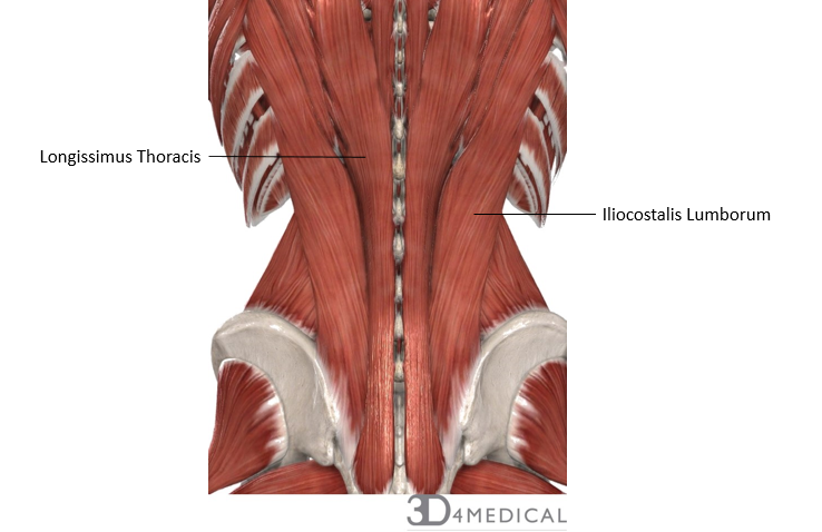
The muscles of the lower back help stabilize, rotate, flex, and extend the spinal column, which is a bony tower of 24 vertebrae that gives the body structure and houses the spinal cord.
Support and protect your spine; The anatomy of your back muscles can be complex. This picture also contains humerus, olecranon process of ulna, deep to tendon and so on. The muscles on each side form a trapezoid shape. Muscular system diagram posterior (back) view. Muscle diagrams are a great way to get an overview of all of the muscles within a body region. To learn more about the anatomy of the spine, watch this video. People with back pain people who experience headaches printing for use during doctor visits to communicate information about your symptoms quickly tracking your progress over time related tools: Broadly considered, human muscle—like the muscles of all vertebrates—is often divided into striated muscle, smooth muscle, and cardiac muscle. To see a muscular system picture from the anterior (front) view click here. Muscles of lower back diagram. (the short head of the biceps femoris even. Diagram of neck and back muscles diagram of neck and back muscles diagram of neck and back muscles upper back muscles cephalicvein.
The back comprises the dorsal part of the neck and the torso (dorsal body cavity) from the occipital bone to the top of the tailbone. Lower back muscle diagram anatomy does degenerative disc disease affect the lower back muscle? Start with 5 to 10 minutes of moderate cardio to get your blood pumping and start to awaken your muscles. Diagram of neck and back muscles diagram of neck and back muscles diagram of neck and back muscles upper back muscles cephalicvein. It is opposite from the chest, and the vertebral column runs down the back.
Human Anatomy Body - Human Anatomy for Muscle ... from www.anatomylibrary99.com For example, some muscles located in the chest also help move the shoulders. The muscles on each side form a trapezoid shape. The muscles of the back can be arranged into 3 categories based on their location: The trapezius is a broad, flat and triangular muscle. The back comprises the dorsal part of the neck and the torso (dorsal body cavity) from the occipital bone to the top of the tailbone. This muscular system diagram shows the major muscle groups from the back or posterior view. We hope this picture anatomy of back muscles diagram can help you study and research. Broadly considered, human muscle—like the muscles of all vertebrates—is often divided into striated muscle, smooth muscle, and cardiac muscle.
For more anatomy content please follow us and visit our website:
Superficial back muscles, intermediate back muscles and intrinsic back muscles.the intrinsic muscles are named as such because their embryological development begins in the back, oppose to the superficial and intermediate back muscles which develop elsewhere and are therefore classed as extrinsic muscles. Broadly considered, human muscle—like the muscles of all vertebrates—is often divided into striated muscle, smooth muscle, and cardiac muscle. The part of the nerve that emerges out of the spine is called the nerve root. The anatomy of your back muscles can be complex. Muscles of lower back diagram. Creatine research more than a sports supplement read more…. How many muscles are in the back? Likewise, there are muscles in other parts of the body that help support and move the spine. (the short head of the biceps femoris even. As you can see, there are also have a spine of scapula deltoid, triceps brachii, latissimus dorsi. To learn more about the anatomy of the spine, watch this video. These structures work together to support the body, enable a range of movements, and send messages from the. Muscles of the head and neck anatomy pictures and inform.